Ultrasound Transducer Types and How to Select the Right Transducer
To utilize the full potential of your ultrasound system, you need the right accessories.
Therefore, the correct ultrasound transducer types are the key to the performance of your ultrasound.
In this blog post, we will explain the different ultrasound transducer types and determine the types of examinations you can use them for.
In the end, we will offer some good points you should keep in mind when you are purchasing transducers.
But first of all
– What is an ultrasound transducer and what does it do?
An ultrasound transducer, also called a probe, is a device that produces sound waves that bounce off body tissues and make echoes.
The transducer also receives the echoes and sends them to a computer that uses them to create an image called sonogram.
Moreover, the essential element of each ultrasound transducer is a piezoelectric crystal.
It serves to generate as well as receive ultrasound waves. Sadly, the medical imaging industry was using the same piezoelectric material for over 40 years.
Up until a few years ago.
Then, a new type of crystal material and ultrasound probe technology appeared. That meant a dramatic improvement in image quality. You can read more about the technology in our blog post – Ultrasound Probe Technology.
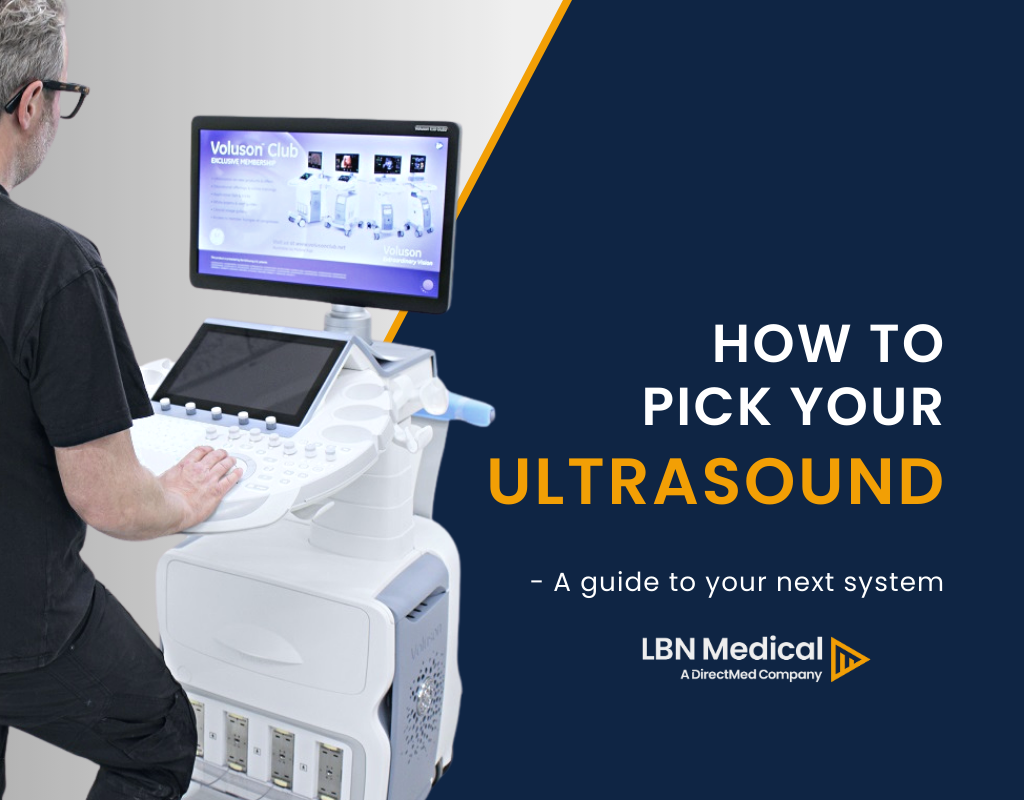
If you would like to learn more about ultrasounds to prepare yourself for choosing a model, you can also sign up to receive our new e-book: “How to pick an ultrasound”
And you become part of our Ultrasound mail course as well. It only costs your e-mail address. You can also read our Guide to Types of Ultrasound Machines.
Get the Ultrasound E-book
Ultrasound Transducer Types
You can find ultrasound transducers in different shapes, sizes, and have diverse features. That is because you need different specifications for maintaining image quality across different parts of the body.
Transducers can be either passed over the surface of the body – external transducers or can be inserted into an orifice, such as the rectum or vagina – these are internal transducers.
Any more differences?
Yes!
The ultrasound transducers differ in construction based on:
- Piezoelectric crystal arrangement
- Aperture (footprint)
- Frequency
Let us go through one at a time.
Footprint also called the aperture, is the part of the probe that is in contact with the body and comes in different shapes and sizes. The footprint is linked to the piezoelectric crystal arrangement, for instance, with linear and convex probes.
The piezoelectric crystal arrangement is the part that obtains the image. Therefore, it affects the footprint but also decides the shape of the ultrasound beam.
Frequency means the frequency of the sound waves emitted from the probe. Generally, higher frequencies offer better image quality, but not as deep penetration compared to lower frequencies.
Below we list the three most common ultrasound transducer types – linear, convex (standard or micro-convex), and phased array. Furthermore, we included other transducers that are available on the market, those are pencil and endocavitary probes.
Linear Transducers
So, what features are typical for the linear transducer (such as GE 9L)?
Firstly, the piezoelectric crystal arrangement is linear, the shape of the beam is rectangular, and the near-field resolution is good.
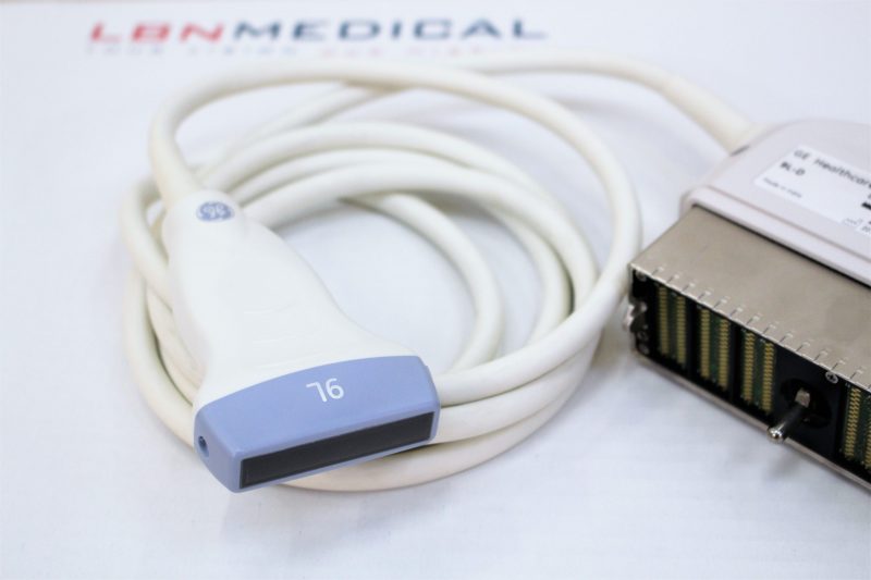
Secondly, the footprint, frequency, and applications of the linear transducer depend on whether the product is for 2D or 3D imaging.
Furthermore, the linear transducer for 2D imaging has a wide footprint and its central frequency is 2.5Mhz – 12Mhz.
You can use this transducer for various applications, for instance:
- Vascular examination
- Venipuncture, blood vessel visualization
- Breast
- Small parts
- Thyroid
- Tendon, arthrogenous
- Intraoperative, laparoscopy
- The thickness measurement of body fat and musculus for daily health care check and locomotive syndrome check
- Photoacoustic imaging, ultrasonic velocity change imaging
The linear transducer for 3D imaging has a wide footprint and a central frequency of 7.5Mhz – 11Mhz.
What can you use this transducer for?
- Breast
- Thyroid
- Arteria carotis of vascular application
Convex Transducers
Another of the ultrasound transducer types is the convex ultrasound transducer, such as GE C1-6, and is also called the curved transducer because the piezoelectric crystal arrangement is curvilinear.
Moreover, the beam shape is convex and the transducer is good for in-depth examinations.
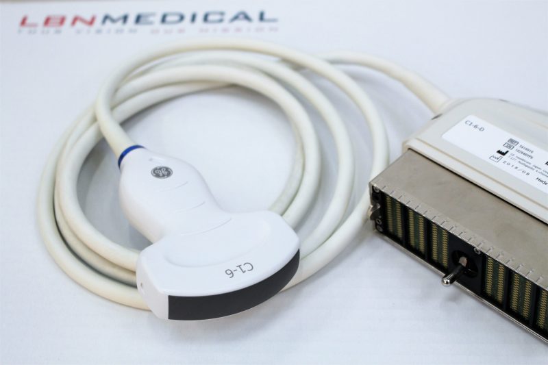
Even though the image resolution decreases when the depth increases. The footprint, frequency, and applications also depend on whether the product is for 2D or 3D imaging.
For example, the convex transducer for 2D imaging has a wide footprint and its central frequency is 2.5MHz – 7.5MHz.
You can use it for examinations such as:
- Abdominal
- Vascular
- Nerve
- Musculoskeletal
- OB/GYN
- Transvaginal and transrectal
- Diagnosis of organs
The convex transducer for 3D imaging has a wide field of view and a central frequency of 3.5MHz – 6.5MHz.
You can use it for abdominal examinations.
In addition to the convex transducers, there is a subtype called micro convex. It has a much smaller footprint and typically, physicians would use it in neonatal and pediatrics applications.
Phased Array Transducers
This transducer is named after the piezoelectric crystal arrangement which is called phased array and it is the most commonly used crystal.
Phased Array transducer has a small footprint and low frequency (its central frequency is 2Mhz – 7.5Mhz).
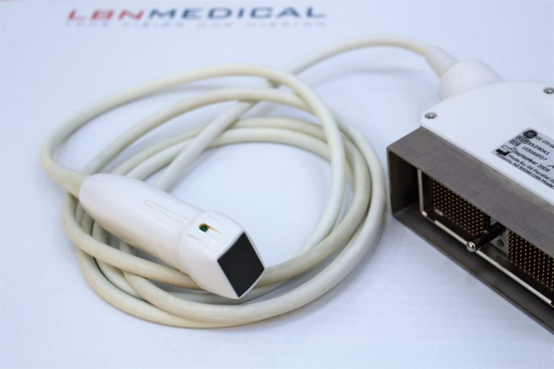
The beam point is narrow but it expands depending on the applied frequency. Furthermore, the beam shape is almost triangular and the near-field resolution is poor.
What can you use the Phased Array transducer for?
- Cardiac examinations, including Transesophagealexaminations
- Abdominal examinations
- Brain examinations
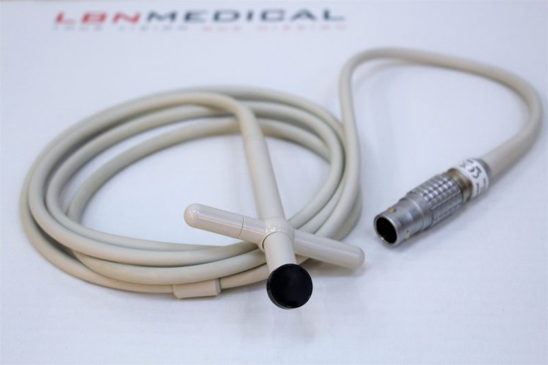
Pencil Transducers
Also called CW Doppler probes, are utilized to measure blood flow and speed of sound in blood.
This probe has a small footprint and uses low frequency (typically 2Mhz– 8Mhz).
Endocavitary Transducers
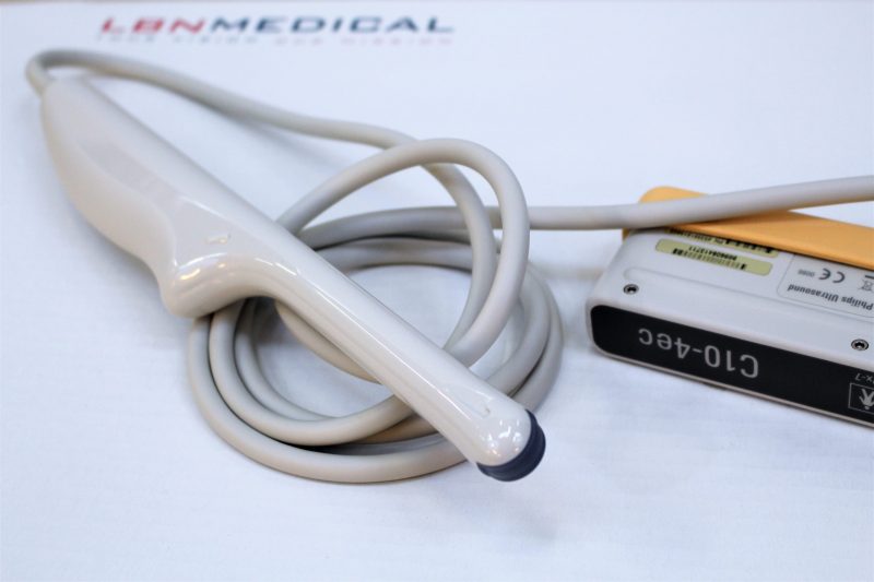
Furthermore, on the list of ultrasound transducer types, there is the endocavitary ultrasound transducer type. These probes provide you with the opportunity to perform internal examinations of the patient. Therefore, they are designed to fit in specific body orifices.
The endocavitary transducers include endovaginal, endorectal, and endocavity transducers (for example, the Philips C10-4EC endocavitary probe below).
Typically, they have small footprints and the frequency varies in the range of 3.5Mhz – 11.5Mhz.
There is also the transesophageal (TEE) probe. As well as the previously mentioned probes, it has a small footprint and is used for internal examinations.
It is often employed in cardiology to obtain a better image of the heart through the oesophagus. The frequency is middle, in the range of 3Mhz – 10Mhz.
Moreover, there are several probes designed for surgical use, such as laparoscopic probes.
To get a quick overview of the different ultrasound transducer types and their applications, see the table below:

Tips You Should Follow When Buying an Ultrasound Transducer
Now, you should be aware of the most common ultrasound transducer types. And we have a few tips that you should follow when purchasing ultrasound transducers:
- Make sure to double-check that the probe you are about to buy is compatible with the system you own – you can use a probe guide, or ask our sales team.
- Penetration depth is better at a low frequency (between 2.5 and 7.5Mhz) but a disadvantage of the low frequency is a lower image quality
- The higher the frequency (above 7.5Mhz), the lower is the depth of penetration, however, you get better quality images close to the surface (7.5MHz = 20 cm).
Be cautious!
- A black line on the screen of the ultrasound system will most likely mean that the transducer has a dead crystal inside.
- A shadow on the screen of the ultrasound system could indicate a weak crystal inside the transducer that does not produce the necessary vibration.
How Should You Treat Your Transducer?
Finally, remember that the transducer is a very important, and also a very expensive element of an ultrasound. Therefore, after you have purchased it, remember that no matter the ultrasound transducer type, you should use it with caution, which means:
- Do not throw, drop, or knock the transducer
- Be careful not to damage the duct of the transducer
- Wipe the gel from the transducer after each use
- Do not sluice with alcohol-based confections
To learn more about how to protect your ultrasound probes and what the most common defects are, check our blog post that explains this topic in more depth.
To conclude, we hope that after reading this article, you have a clear image of ultrasound transducer types. And that you will be more prepared the next time you are purchasing probes.
If you have any more questions about transducers or used ultrasound machines, do not hesitate to contact our sales department at sales@lbnmedical.com or via phone +45 96 886 500.
Would you like to purchase a probe but you don’t feel like sending us an email or calling us?
Just fill in this form with your request. We will get back to you as soon as possible!
Also, check our inventory of ultrasound machine parts.
What is next?
If you would like to learn even more about ultrasounds, you can sign up below to receive an e-book: ‘How to pick your next ultrasound’ and become part of our ultrasound e-mail course.
In multiple e-mails, this course will guide you through several themes related to your next ultrasound purchase.
